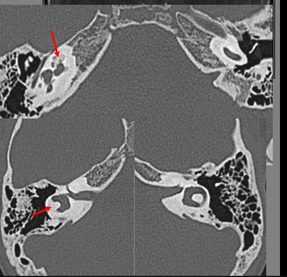
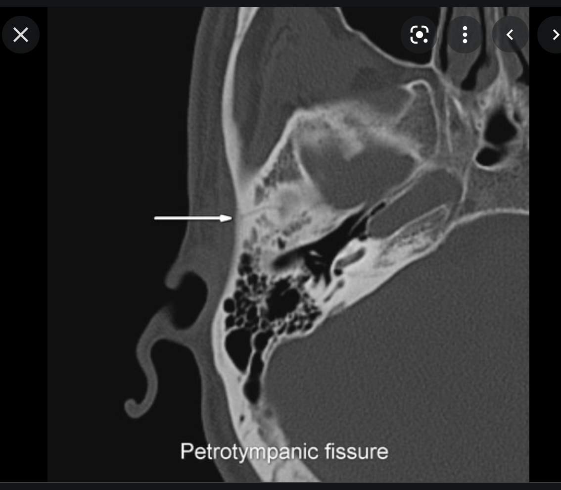
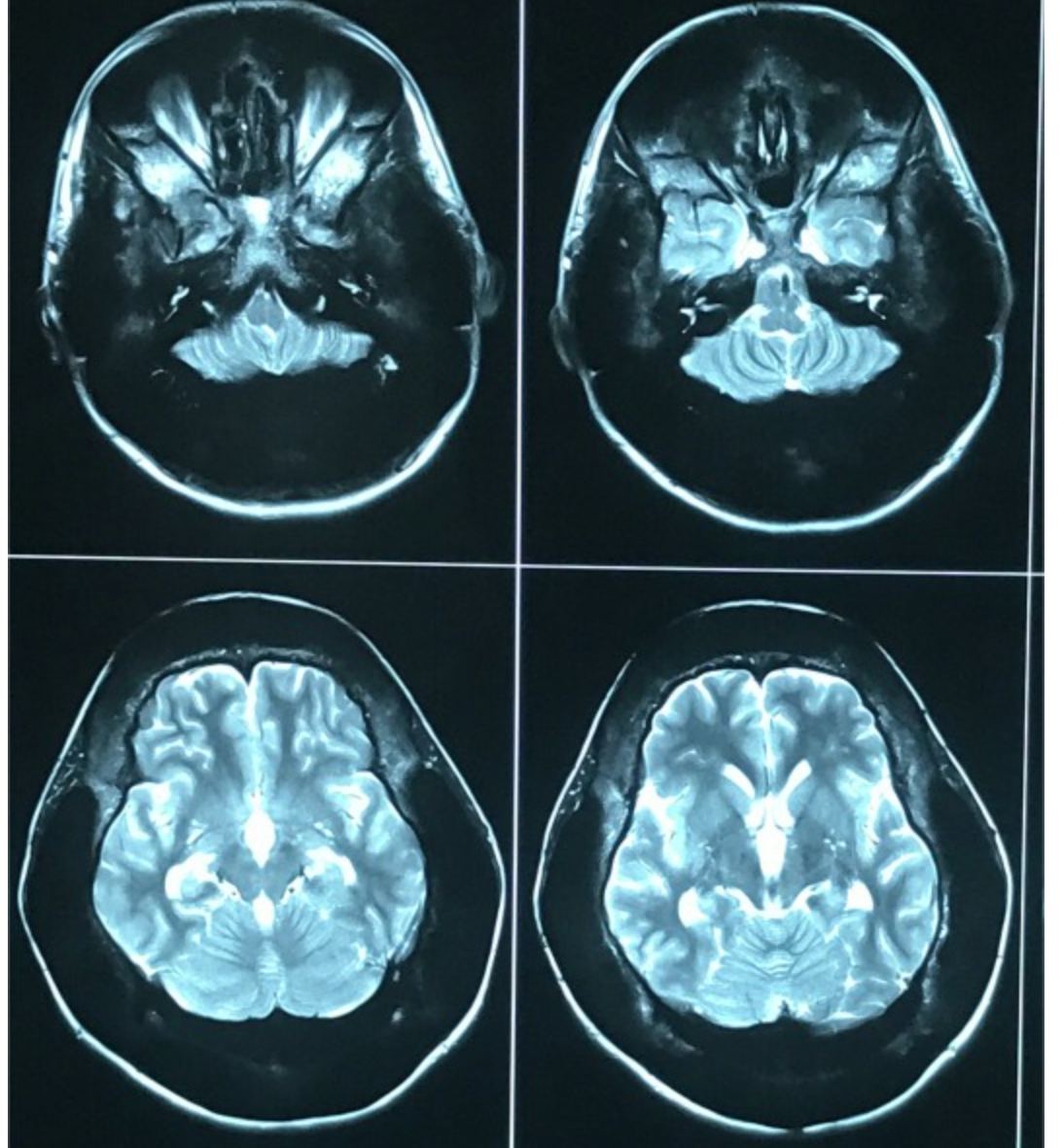
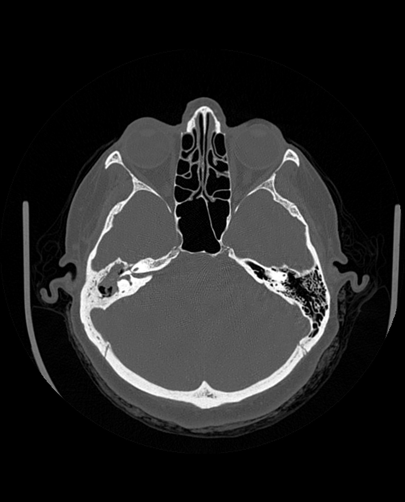
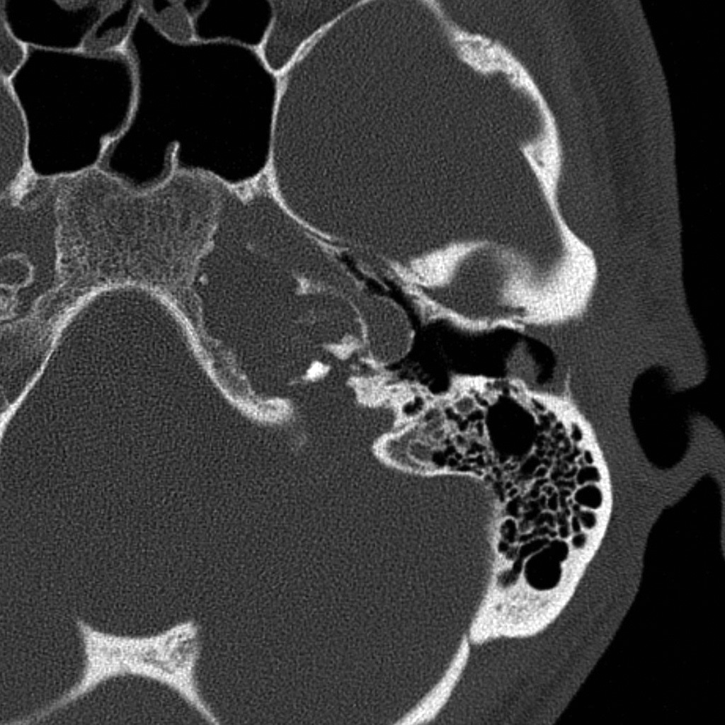
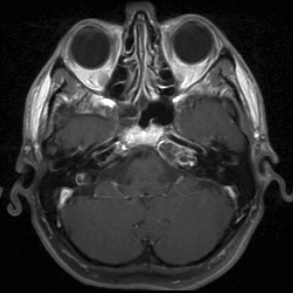
cochlear aplasia

The labyrinth is abnormal with the cochlear absent (cochlear aplasia). The vestibule, semicircular canals and ossicles are present.
Cochlear aplasia, or complete absence of the cochlea is a rare anomaly which accounts for only 3% of cochlear malformations.
Radiographic features
- complete absence of the cochlea. Dense otic bone is seen at the anatomical site of the cochlea 2
- cochlear nerve canal and cochlear nerve are absent
- cochlear promontory is hypoplastic and flattened
- the vestibule and semicircular canals are often malformed, stunted, dilated but may be normal
- vestibular aqueduct is normal
- internal auditory canal usually hypoplastic
- facial nerve canal usually anomalous showing obtuse angle anterior genu
- middle ear is usually normal sized with normal ossicles
- oval window usually normal but may be atretic
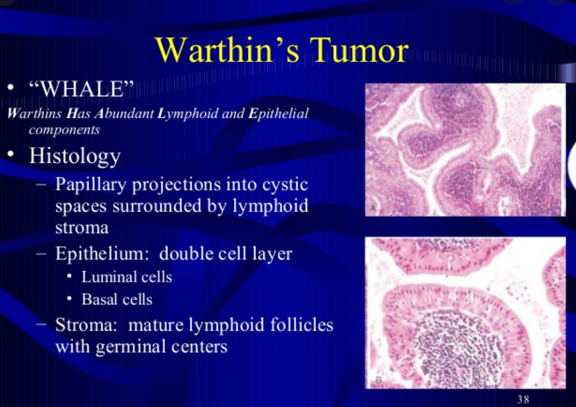
Which benign H+N tumour is comprised of Epithelial and lymphoid cells.
Warthins Tumour
what is this?
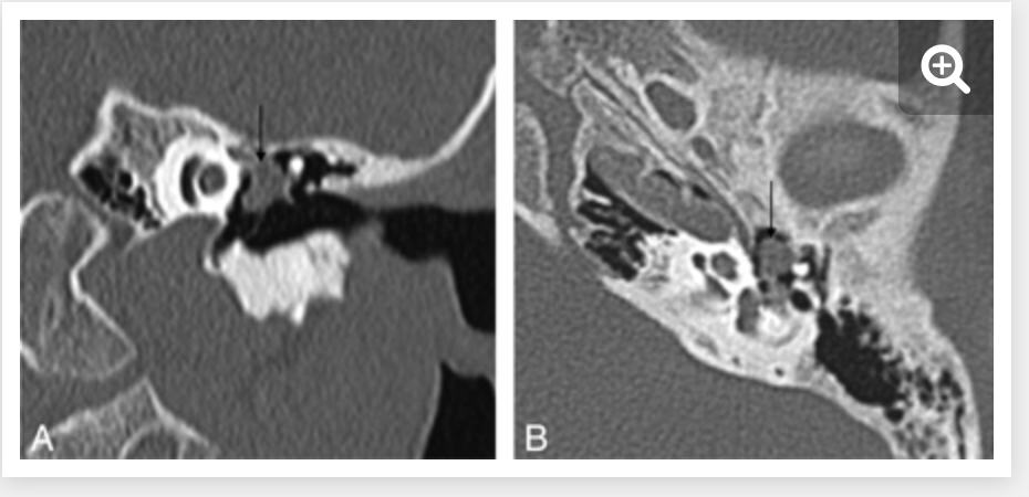
- Congenital cholesteatoma
- 2% of temporal bone cholesteatomas
- can effect the middle ear, EAC, mastoid or petrous bone, or labyrinthe
- Most common location is the anterosuperior portion of the middle ear near the eustachian tube or stapes.
- CLinical
- young pt with normal mastoid pneumatization and without hx of chronic ear infections
- Histo
- squamous cell lining
- keratin debris
- cholesterol
rad features of this tumour
Typical location
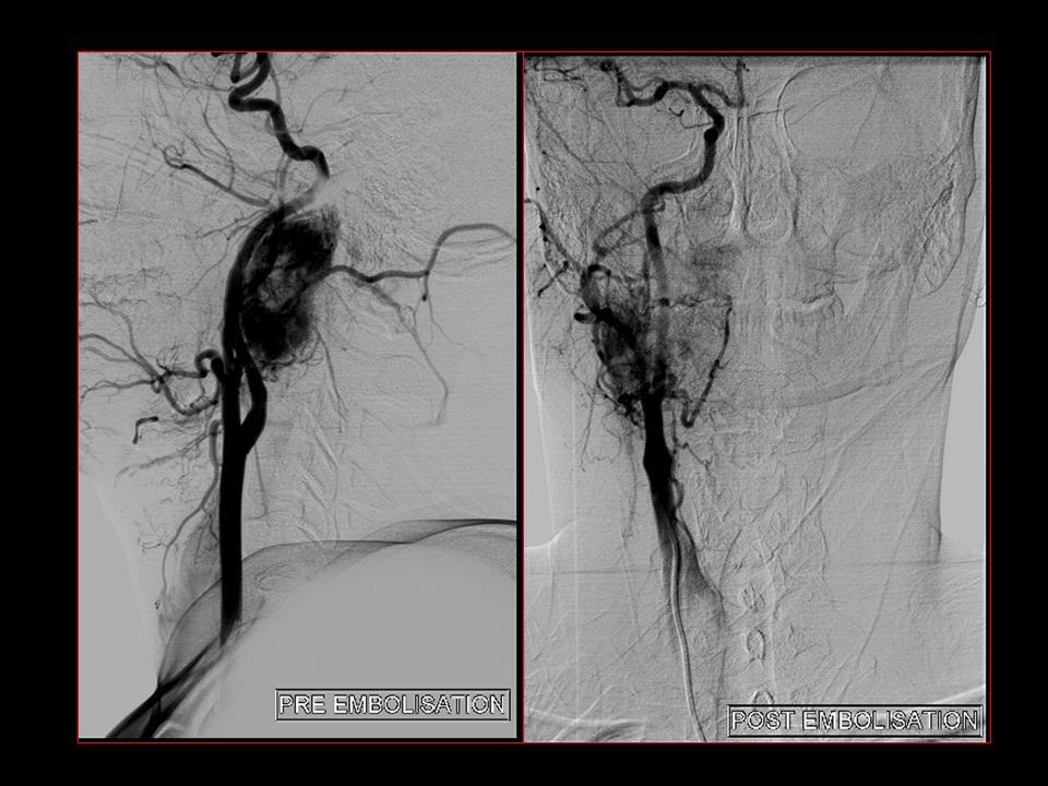
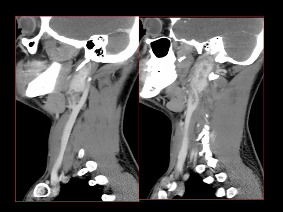
- Rad Features of Glomus Vagale Paraganglioma tumours
- arises from paraganglia in the nodose ganglion of the vagus nerve in the nasopharyngeal carotid (or post-styloid parapharyngeal) space.
- displaces carotid anteromedially
- displaces jugular vein posterolateral and
- Does not splay the carotid bifurcation.
Glomus vagale tumours are paragangliomas that occur along the path of the vagus nerve (CN X). They are a subset of extra-adrenal neuroendocrine tumours that are derived from the nonchromaffin paraganglion cells.
Clinical presentation
Typically presents as a painless mass behind the carotid artery. Vocal cord paralysis is a relatively frequent finding (~47%) 3.
Location
Although they could occur at a similar position to carotid body tumours they tend to be more rostral in location and do not widen the carotid bifurcation. They displace the internal and external carotid arteries anteriorly, and the internal jugular vein posteriorly 1.
Case courtesy of Dr Ashwin M Polnaya, Radiopaedia.org, rID: 5926
Contents of Foramen Ovale
V3
accessory meningeal artery
Otic ganglion

Contents of Foramen Rotundum
V2
artery of foramen rotundum
emissary veins
What are Glomus tumours?
AKA
What do they arise from?
- Glomus tumors
- aka
- chemodectomas
- arise from chemoreceptor cells in multiple sites in the head and neck
- The majority are benign and approx 10% are multiple
- therefore it is important to check other common locations in the H+N during imaging
- GLomus tympanicum represents the. most common middle ear tumor
*
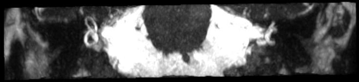
- Labyrinthitis ossificans
- ossification of the membranous labyrinth as a sequela of previous infections, inflammatory, traumatic or surgical injury to the middle ear.
- Seen as ossification within the membranous labyrinth on HRCT and foci of low signal on T2 with the otherwise high signal fluid of the membranous labyrinth
- Radiographic features
The scala tympani of the basal turn of the cochlea is the most commonly affected site 10.
CT
High-density bone deposition within the membranous labyrinth:
mild disease: hazy increase in density within fluid spaces of the membranous labyrinth
moderate disease: focal areas of bony encroachment on fluid spaces of the membranous labyrinth
severe disease: membranous labyrinth completely obliterated by bone replacing fluid spaces
MRI
loss of normal high signal of fluid within the membranous labyrinth is seen on heavily T2 weighted images (as low signal intensity foci in the labyrinth)
What is this?

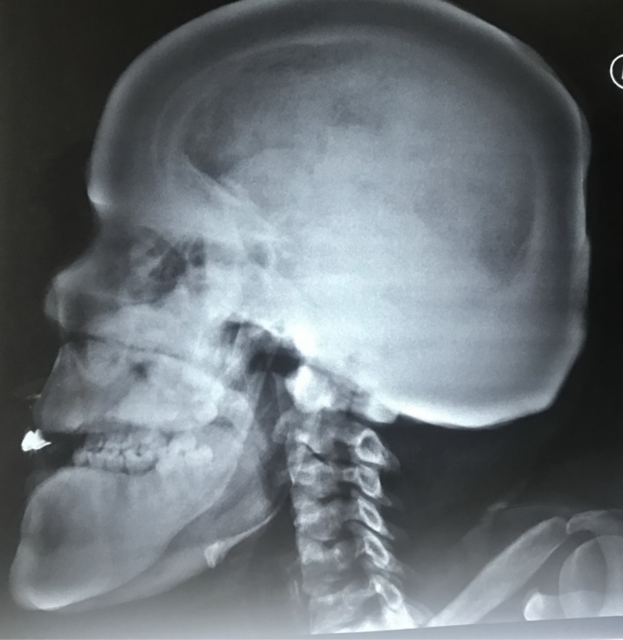
- Klippel Fiel Syndrome:
- Cervical fusion
- short neck
- low posterior hairline
- limited cervical motion
Case courtesy of Dr Mohammad A. ElBeialy, Radiopaedia.org, rID: 23924
what does the anterior skull base consist of?

Broadly consists of floor of the anterior cranial foss and the roof of thenose, ethmoid air cells and orbits.

what is the pterygopalatine fossa
- an important space and potential rout of spread of disease in the deep face
- Contents
- Pterygopalatine ganglion
- Maxilary Nerve (V2) via foramen rotundum
- distal internal maxillary artery (via pterygomaxillary fissure)

Contents of the Optic Canal
3
- Optic Nerve
- Ohthalmic artery
- opthalmic veins

What are the different types of Glomus tumours?
4
types
- glomus jugulara:
- origin at the jugular bulb
- more common
- Glomus tympanicum
- arises from paraganglia along the inferior tympanic nerve (Jacobson nerve) frequently on the cochlear promontory
- Glomus vagale
- carotid body tumour
what does the central skull base consist of?
- floor of the middle crainal fossa,
- roof of the sphenoid sinus
- greater wing of sphenoid

what are the 2 types of fractures of the temporal bone?
which is more common?
Typically involves?
Which one involves the ossicles?
Which one involves the TM?
which is more likely to cause facial paralysis?
- Longitudinal Fractures
- red line
- 80% (more common)
- parallel to long axis
- Typically involves the middle ear
- The labrynth is typically spared
- The ossciles are usually involved (conductive hearing loss)
- The TM is is involved
- Facial paralysis 20%
- Transverse fractures
- Yellow line
- 20% (less common)
- Perpendicular to long axis
- Typially involves the inner ear
- The labyrinth is commonly involved
- the ossicles and TM are frequently spared
- Facial paralysis 50%
- A more clinically relevant classification may be otic capsule violating vs sparing.
- Otic capsule involvement increases the risk of sensorineural hearing loss, 7th CN palsy and CSF leak
- Fractures may also be oblique: ie mixed features of longitunial and transverse fractures, cross the petrotympanic fissure.
- Otosclerosis, also known as otospongiosis, is a primary osteodystrophy of the otic capsule (bony labyrinth of the inner ear). It is one of the leading causes of deafness in adults.
Terminology
The term otosclerosis is somewhat of a misnomer. Much of the clinical course is characterised by lucent rather than sclerotic bony changes and hence it is more appropriately known as otospongiosis which is a term preferred by many head and neck radiologists.
Contents of the vidian canal
- aka pterygoid canal
- vidian nerve
- vidian artery
- vidian vein
- connects PPF anteriorly to the foramen lacerum posteriorly

Petrous malformations a/w recurrent meningitis
- The presence of a fistula in the petrous portion of the temporal bone may lead to otorrhea, pneumocephalus, meningitis or abscess
- Acquired lesion of the petrous apex can potentially have similar complications depending on extent and violation of adjacent structures.

Craniodiaphyseal Dysplasia
Craniodiaphyseal dysplasia is a very rare autosomal recessive disorder which is typically presented in infancy and characterised by severe form of bone dysplasia, massive bone sclerosis and hyperostosis. This process of bone changes characteristically affects the facial bones resulting in severe facial deformity.
https://www.sciencedirect.com/science/article/pii/S2214541919300392
What are the complications of Acquired Cholesteatoma?
What complication/s are demonstrated here?
- Labyrinthine fistula
- dehiscence of semicircular canals
- usually the lateral one.
- Facial nerve paralysis
- 2ndary to involvement of the facial nerve canal
- Invasion of the tegmen tympani, petrous apex or sigmoid plate
- Automastoidectomy
Automastoidectomy denotes extensive bone destruction of the mastoid mimicking the appearance of surgery (mastoidectomy), most often caused by cholesteatoma.
Spontaneous evacuation of cholesteatoma can be seen with automastoidectomy 1. In these circumstances, it is often referred to as mural cholesteatoma or unusual cholesteatoma shell, as there is no residual soft tissue mass 2.
Automastoidectomy has also been reported with keratosis obturans 3.
https://radiopaedia.org/cases/automastoidectomy-with-labyrinthine-fistula
Automastoidectomy refers to extensive bony destruction of the mastoid forming one cavity resembling operative mastoidectomy. Cholesteatoma is the most common cause and this subtype is known as “mural cholesteatoma”.
Labyrinthine fistula refers to abnormal communication between inner ear perilymph and middle ear cavity on top of erosion or fracture of the bony labyrinth.

Treacher Collins syndrome
- atretic external auditory canal with hypoplastic middle ear and absent ossicles
- midline cleft palate
- underdeveloped zygomatic and pterygoid part
- hypoplastic mandible and maxilla
- anterior concavity of mandible
- hypoplastic left parotid
- parotid duct calculi
1 case question available
Case Discussion
In Treacher Collins syndrome, 1st and 2nd branchial arch structures are affected but the inner ear structures are normal.
Treacher colins Syndrome

- atretic external auditory canal with hypoplastic middle ear and absent ossicles
- midline cleft palate
- underdeveloped zygomatic and pterygoid part
- hypoplastic mandible and maxilla
- anterior concavity of mandible
- hypoplastic left parotid
- parotid duct calculi
1 case question available
Case Discussion
In Treacher Collins syndrome, 1st and 2nd branchial arch structures are affected but the inner ear structures are normal.

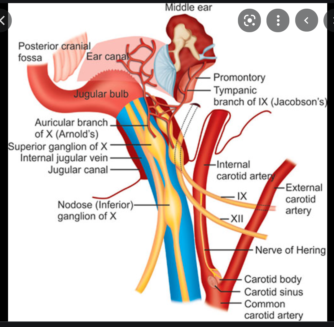
Gradenigo syndrome consists of the triad of:
petrous apicitis
abducens nerve palsy, secondary to involvement of the nerve as it passes through Dorello canal
retro-orbital pain, or pain in the cutaneous distribution of the frontal and maxillary divisions of the trigeminal nerve, due to extension of inflammation into Meckel cave
Pathology
Common pathogens are Pseudomonas and Enterococcus spp.
History and etymology
It was first described in 1907 by Giuseppe Conte Gradenigo (1859-1926), Italian otolaryngologist 2,3.
3 Places where lesions of the anterior skull base can arise from
- Centered in the bone
- arise in the anterior cranial fossa abvoe
- arise in the sinonasal cavities below
localisation of the lesion centre can help with the differential


-
CHEST IMAGING 1100
-
CHEST IMAGING 2100
-
CHEST IMAGING 343
-
CHEST IMAGING 4 (TUMOURS)81
-
CHEST IMAGING 5 PATHOLOGY2
-
CARDIAC IMAGING 1100
-
CARDIAC IMAGING 2100
-
CARDIAC IMAGING 3100
-
CARDIAC IMAGING 443
-
GIT 1101
-
GIT 2100
-
GIT 3100
-
GIT 4102
-
Hepatobilary155
-
Biliary System76
-
Pancreas66
-
Spleen24
-
Adrenal Glands70
-
GENITOURINARY IMAGING 197
-
GENITOURINARY IMAGING 2100
-
GENITOURINARY IMAGING 3100
-
GENITOURINARY IMAGING 4100
-
GENITOURINARY IMAGING 545
-
RETROPERITONEUM33
-
Male Pelvis10
-
GIT PATHOLOGY31
-
Skeletal Dysplasias18
-
MSK104
-
MSK Crack the Core70
-
MSK 295
-
MSK 3100
-
Neuro100
-
Neuro 222
-
NEURO 375
-
Head and Neck 1100
-
Head and Neck 2100
-
Head and Neck 3100
-
Head and Neck 4100
-
Head and Neck 555
-
DDX Head and Neck35
-
Vascular10
-
IR30
-
BREAST IMAGING52
-
OBSTETRICS17
-
GYNAECOLOGY40
-
PAEDIATRICS 196
-
PAEDIATRICS 297
-
PAEDIATRICS 395
-
Nuclear Medicine 134
-
PET CT16
-
Syndromes94
-
HAEMATOLOGY6
-
PATHOLOGY 141
-
Crack the core WHen I Say you say...489
-
Physics31
-
crack the core exam case companion18
-
EPONYMOUS Diseases/signs22
-
What the F&^# is that word?10
-
Radiology Signs25
-
Mnemonics36
-
GIT Pathology1
-
NEURO MRI PHYSICS14
-
GREAT CHEST XRAY CASES1
-
THIS PATIENT IS TYPICAL OF X CONDITION2
