
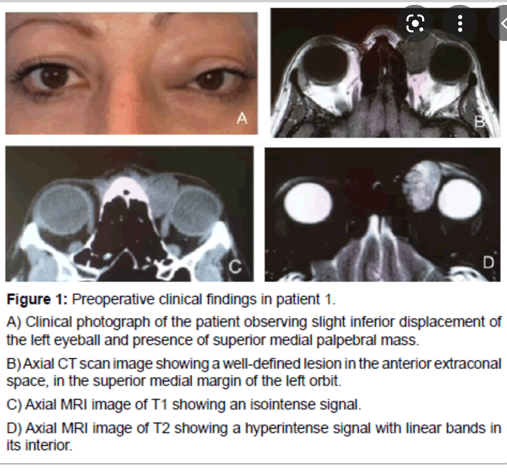
Anterior skull base Mucocele
- expanded sinuse from chronic obstruction
- variable signal depending on protein content of secretions
- No solid enhancement
- can see mucosal enhancement around the margins
- Non-agressive with bone remoddeling
- MRI highly accurate for distinguishing higher density/proteinaceaus secreations that are equivocal on CT from Solid masses
- DDX
- mucus retention cyst
- does not completely fill the sinus
- no bony expansion
- paranasal sinus carcinoma (generally isointense or of intermediate intensity on MR imaging sequences) 5
- aspergillus sinusitis
- dermoid cyst
- osteoma (rare) 6
- mucus retention cyst

PNS can be antegrade and retrograde
True or false?
true.
What is PHPV associated with?

- Other ocular dysplasias
- Norrie disease
- seizures
- deafness
- low IQ
Rad Features of Optic Nerve Meningioma

- Mass
- tubular 65%
- Exophytic/eccentric 25%
- Fusiform/surrounding the optic nerve 10%
- Calcification common
- Enhancement
- intense contrast enhancement
- Linear bands of enhancement (nerve within tumour) Tram track sign
- Sphenoid bone +/- optical canal hyperostosis in advanced tumours

Clinical symptoms of Optic nerve glioma
- Loss of vision
- proptosis (in bulky tumours)
- Occur most commonly in the 1st decade of life
- May be bilateral in NF1
Orbital hemangiopericytoma
Orbital hemangiopericytoma
- uncommon
- slow growing vascular neoplasm
- can be benign or malignant
- well-circumscribed or invasive when high grade
- may remodel or erode bone
- avidly enhancing
- flow voids common on T2
Buphthalmos

Buphthalmos
Buphthalmos:
- congenital glaucoma, anterior ocular chamber drainage problem
- enlarged globe
- increased depth of anterior chamber
Causes of Optic Neuritis
- Idiopathic
- MS
- Neuromyelitis Optica (Devic syndrome)
- Acute demyelinating encephalomyelitis (ADEM)
- Paediatric Optic Neuritis (may follow viral illness or vaccination, ADEM)

- Anterior skull base Meningocele/encephalocele
- extension of meninges +/- brain parenchyma through skull base defect into nasal cavity or ethmoidal labyrinth.
- CT can show bone defect
- high res MRI for confirmation
- Heavily T2 high rest sequences can be helpful for clear demonstration of extension of meninges and or brain through defect.
- https://thejns.org/focus/view/journals/neurosurg-focus/32/6/2012.3.focus1267.xml

what does ocular melanoma arise from?
- arises from pigmented choroidal layer
- retinal detachment is common
Ecchordosis Physaliphora

Ecchordosis Physaliphora
Cystic lesion in the prepontine cistern without enhancement or restricted diffusion extending into bone.
Absence of enhancement or restricted diffusion together with non-aggressive bony margins in this location is typical of a retroclival ecchordosis physaliphora, a notochord remnant.
Ecchordosis physaliphora is a congenital benign hamartomatous lesion derived from notochord remnants, usually located in the retroclival prepontine region, but can be found anywhere from the skull base to the sacrum.
Terminology
There has been some controversy as to whether intradural chordoma and large ecchordosis physaliphora are different entities. Some authors (such as Wolfe et al.) proposed the name ‘intradural chordoma’ for all intradural notochordal remnant lesions 8. Others (such as Rodriguez et al.) proposed that all intradural notochordal remnant lesions should be called ecchordosis physaliphora, until chordoma are pathologically proven to arise from the intradural compartment 9. However, they are currently considered distinct pathologies with a common origin.
Clinical presentation
Unlike chordomas which are often symptomatic due to brainstem or cranial nerve compression, patients with ecchordosis physaliphora are usually asymptomatic. They are found in ~2% of autopsies 1.
Pathology
Ecchordosis physaliphora arise from remaining notochord cells along the axis of the spine after embryogenesis. Unfortunately, ecchordosis physaliphora and chordoma are histologically indistinguishable, other than by examining the margins, the latter demonstrating infiltrative growth.
Central skull base Aneurysms

Central skull base Aneurysms
- Cavernous or other ICA aneurysms
- Rounded ‘mass’
- low signal
- flow void on MRI MUST RECOGNISE
- Pulsation artifact in phase enconding direction
- CT or MRI angio to confirm
Rhabdomyosarcoma
Rhabdomyosarcoma
- Most common malignant orbital tumour in childhood.
- mean age 7 years
- Large aggressive soft tissue mass
- intraconal or extraconal
- mets to lung and cervical nodes
- Rhabdomyosarcomas of the orbit account for approximately 10-20% of all rhabdomyosarcomas and are usually found in children.
- Epidemiology
- As with other locations, rhabdomyosarcomas in the orbit are overrepresented in males, and in Caucasians.
- They typically occur in children below the age of 15 years.
- Clinical presentation
- Clinical presentation is typically with a rapidly enlarging mass, often in the upper inner quadrant 1.
- It is usually painless but causes proptosis and diplopia.
- Often the mass invades the eyelid causing marked oedema 1.
- Pathology
- The vast majority of orbital rhabdomyosarcomas are of the embryonal subtype 1,3.
- Contrary to early belief, these tumours do not arise from the extraocular muscles, but rather develop from primitive mesenchymal cells that go on to differentiate into striated muscle cells 3.
- The vast majority of orbital rhabdomyosarcomas are of the embryonal subtype 1,3.
- Histologic subtypes:
- embryonal
- alveolar
- mixed
- Radiographic features
- CT and MRI are the modalities of choice for assessment of these masses, and to delineate adjacent structures.
- It is important to report the location of the tumour epicentre as there is a correlation between location and histology:
- embryonal subtype more frequently arises in the superior orbit, whereas
- alveolar subtype is more common in the interior orbit 3.
What are the radiographic findings of ocular melanoma?
- Thickening or irregularity of choroid
- localised
- polypoid
- flat
- exophytic biconvex mass lesion
- usually initlateral posterior location
- retinal detachment common
- contrast enhancement
- MRI
- T1 bright
- T1 low
*
WHICH nerves does PNS most often occur?
PNS (perineural Spread) most commonly occurs along CN V and VIII branches
Complications of Optic Neuritis
40-60% of patients ultimately develop MS
70-90% of MS patients develop optic neuritis

- Meningioma
- occasionally lesions can be infiltrative, extend to extracranial spaces such as PPF and infratemporal fossa or have intraosseous component
Figure 2: Medial sphenoid wing meningiomas can present different set of technical challenges based on their involvement of the medial neurovascular structures and the encasement of the carotid artery’s perforating vessels. A medial sphenoid wing meningioma with minimal medial extension is shown (upper images). The Sylvian middle cerebral artery branches drape over the superior pole of the tumor. A more true medial sphenoid wing/clinoidal meningioma with significant medial extension and encasement of the ICA is also included (lower images).
Orbital Infantile Hemangioma Associated syndrome

PHACES syndrome
Posterior fossa malformation
Hemangioma
Arterial anomalies
coarct/cardiac anomalies
Eye anomalies
Sternal clefting/supraumbilical raphe

Sinonasal melanoma

- Sinonasal mucosal melanoma (SNMM) is a very rare and unique subtype of malignant melanoma.
- ON MRI: lesions may have intrinsically high signal on T1 and low signal on T2 - paramagnetic properties of melanin
- Epidemiology
- SNMMs account for ~1% of malignant melanomas and <4% of head and neck cancers 1,2.
- They affect older patients (60-90 years old) 2.
- There is a higher incidence in Japan 5.
- Clinical presentation
- Headache and visual symptoms are common.
- Pathology
- Typically SNMMs are an expansile mass centred within the nasal cavity (more common) or the paranasal sinuses.
- Invasion is common, in particular to the orbits, base of the skull, intracranially, or nasopharynx.
- Hepatic metastases are common 4.
- Staging
- Mucosal melanoma of the head and neck, including sinonasal mucosal melanoma, is staged according to the American Joint Committee on Cancer TNM system.
- Radiographic features
- Radiographic features of SNMM are variable, especially on MRI, due to varying amounts of melanin, with up to one-third of cases being amelanotic 3.
- CT
- polypoid or mass-like
- bony remodelling +/- erosion commonly present
- strongly contrast enhancing 5
- MRISignal characteristics
- T1: homogeneous T1 signal
- high T1 signal may be seen secondary to haemorrhage or melanin 5
- T2: low signal
- T1C+: moderate homogeneous or heterogeneous enhancement
- Metastases return the same signal characteristics as the primary lesion.
- T1: homogeneous T1 signal
- Treatment and prognosis
- SNMM is aggressive and carries a poor prognosis with a five-year survival rate of ~30% 3.
- Differential diagnosis
- As a general differential in the sinonasal region consider other tumours such as:
- sinonasal undifferentiated carcinoma
- As a broader differential in the nasopharyngeal region, consider other nasopharyngeal tumours such as:
- As a general differential in the sinonasal region consider other tumours such as:

Central skull base Basal cephalocele

Central skull base Basal cephalocele
FIGURE 31-5 Basal encephalocele.A, A sagittal T1-weighted image shows callosal agenesis with a tiny lipoma (arrow). A large defect in the basisphenoid is seen. Note the apparent absence of the pituitary, floor of the third ventricle, and optic pathways. B, A high-resolution sagittal T2-weighted image shows the pituitary-hypothalamic structures (arrow) and optic pathways are contained within the encephalocele.
Anterior skull base Fibrous dysplasia

Anterior skull base Fibrous dysplasia
- expansile developmental bone lesion
- classically ground glass matrix
- varies depending on amount of firbous and ossified components.
- enhancing lesion can be deceptively aggressive on MRI (suspect if very low signal on T1 and T2
- obtain CT to demonstrate typical characteristics
Central skull base Persistent craniopharyngeal canal

Central skull base Persistent craniopharyngeal canal
- developmental anomaly resulting in a persistent tract from nasopharynx to pituitary fossa
- a smoothly marginated midline canal between presphenoid and basisphenoid
- typically incidental finding but may be a/w pituitary abnormalities, cephaloceles, midline craniofacial anomalies or rarely even tumours arising from tissue within the canal

Hemangiopericytoma of the anterior skull base

- hypervascular lesion with aggressive growth pattern
- dural based mas that may have internal flow voids
- intensely enhancing
- The magnetic resonance imaging (MRI) with contrast (A) sagittal (B) axial, and (C) coronal views depict a large contrast-enhancing mass arising from the olfactory groove, D: the MRI T2 weighted image demonstrates corkscrew type flow voids (white arrow), which are consistent with a highly vascular neoplasm.
- https://www.cureus.com/articles/9578-hemangiopericytoma-in-the-olfactory-groove-a-rare-and-unusual-presentation


-
CHEST IMAGING 1100
-
CHEST IMAGING 2100
-
CHEST IMAGING 343
-
CHEST IMAGING 4 (TUMOURS)81
-
CHEST IMAGING 5 PATHOLOGY2
-
CARDIAC IMAGING 1100
-
CARDIAC IMAGING 2100
-
CARDIAC IMAGING 3100
-
CARDIAC IMAGING 443
-
GIT 1101
-
GIT 2100
-
GIT 3100
-
GIT 4102
-
Hepatobilary155
-
Biliary System76
-
Pancreas66
-
Spleen24
-
Adrenal Glands70
-
GENITOURINARY IMAGING 197
-
GENITOURINARY IMAGING 2100
-
GENITOURINARY IMAGING 3100
-
GENITOURINARY IMAGING 4100
-
GENITOURINARY IMAGING 545
-
RETROPERITONEUM33
-
Male Pelvis10
-
GIT PATHOLOGY31
-
Skeletal Dysplasias18
-
MSK104
-
MSK Crack the Core70
-
MSK 295
-
MSK 3100
-
Neuro100
-
Neuro 222
-
NEURO 375
-
Head and Neck 1100
-
Head and Neck 2100
-
Head and Neck 3100
-
Head and Neck 4100
-
Head and Neck 555
-
DDX Head and Neck35
-
Vascular10
-
IR30
-
BREAST IMAGING52
-
OBSTETRICS17
-
GYNAECOLOGY40
-
PAEDIATRICS 196
-
PAEDIATRICS 297
-
PAEDIATRICS 395
-
Nuclear Medicine 134
-
PET CT16
-
Syndromes94
-
HAEMATOLOGY6
-
PATHOLOGY 141
-
Crack the core WHen I Say you say...489
-
Physics31
-
crack the core exam case companion18
-
EPONYMOUS Diseases/signs22
-
What the F&^# is that word?10
-
Radiology Signs25
-
Mnemonics36
-
GIT Pathology1
-
NEURO MRI PHYSICS14
-
GREAT CHEST XRAY CASES1
-
THIS PATIENT IS TYPICAL OF X CONDITION2
