Why biopsy the skin?
- Establish a definitive diagnosis that cannot be reached by other, less invasive, testing methods
- Rule out certain conditions
Biopsy sampling do’s?
- Be gentle (no squeeze, scrubbing or clipping)
- Biopsy early
- Collect multiple samples representative of the range of the lesions
- Include crusts
- Biopsy before using anti-inflammatory therapy
- Label samples from different areas
- Submit a complete history, full signalment, precise description, list of differentials and a diagram
Do a review of normal histology of skin?
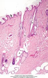
The epidermis (arrow) in haired skin has an undulating surface but lacks rete ridges. The epidermis in haired skin has fewer nucleated cell layers than the epidermis in nonhaired (hairless) skin such as that on the nose and pawpads; thus it is referred to as “thin” skin. Hair follicles (H), apocrine glands (A), and sebaceous glands (S) are present. The haired skin is thickest over the dorsal aspect of the body and on the lateral aspect of the limbs, and it is thinnest on the ventral aspect of the body and the medial aspect of the thighs. H&E stain
Name the different patterns?
Perivascular dermatitis
- Leuokcytes come into the skin, in the dermis (the structure that has blood vessels) – not the epidermis.
Interface dermatitis
- Most of the time hinting at an autoimmune disease
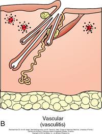
Vasculitis
- Target of inflammation seems to be the vasculature in the dermis. This can be an immune mediated process or a virus; numerous virus that can cause this.
Nodular and/or diffuse dermatitis
Vesicular/pustular dermatitis
- Intraepidermal
- Subepidermal
Folliculitis/furunculosis/sebaceous adenitis
- Inflammation/degeneration centred around hair follicle.
Panniculitis
- Inflammation centred around
Atrophic dermatoses
- Pattern that shows less or no inflammation – more denegerative process, lose skin structures, may be a lack of nutrients etc.
Describe Perivascular dermatitis?
Most common but unfortunately least specific diagnostic pattern
Least specific pattern – it is just inflammation of the skin; leukocytes are sent into the dermis.
- Prominent blood vessels
- Oedema of dermis
- Leukocytes around vessels
- Classified according to depth
- Superficial dermal
- Mid-dermal/perifollicular
- Deep dermal
- Cellular infiltrate varies eg
- Neutrophil
- May be an acute pyoderma etc.
- Neutrophil
- Lymphocytes
- Eosinophil (Type 1 hypersensitivity –parasitic? Allergic?)
- If seen eosinophils, mainly have two pathways: hypersensitivity reaction or parasitic infection.
- Classic examples can be canine atopy, pyoderma, flea bite hypersensitivity…
Describe Interface dermatitis?
- Little bit more serious and specific.
- Band like infiltration of most of the time, lymphocytes that are migrating up into the epidermis. There can be quite cell dense areas, or quite subtle. See degeneration of basal cell layer.
- May see more or less apoptotic keratinocytes – either being attacked or apoptosis is being induced; commonly the case with autoimmune diseases.
- Cell-rich or cell-poor band-like mononuclear infiltrate crossing dermo-epidermal junction
- Hydropic degeneration of basal keratinocytes
- +/- apoptosis (individual cells, mainly in basal layer)
- Pigment incontinence
- Associated with immune-mediated diseases
- Examples for interface dermatitis are dermatomyositis, erythema multiforme , lupoid dermatoses, VKH and many others..
Describe vasculitis?
- Inflammation of blood vessels
- Leukocytes come in and target little vascular structures; may see micro-heamorrhages
- Can involve subcutaneous fat tissue.
- Tight perivascular cuffs of inflammatory cells with degeneration of vascular wall
- Suspect if see microhaemorrhages
- Variety of cell types
- +/-
- Panniculitis
- Dermal necrosis
- Atrophy of hair follicles
- Either primary or secondary to inflammation, infection, drug reactions, neoplasia, vaccination
- Can be difficult to find on sampling – need early lesion. Take multiple samples.
- Examples are dermatomyositis, rabies-vaccine induced panniculitis in dogs, pastern dermatitis in horses… also classical swine fever, malignant cattahal fever
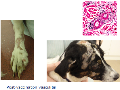
Image showing post vaccination vasculitis. Massive accumulation of Ag.Ab complexes depositing in vascular wall and activating complement cascade = inflammation.
Describe Nodular and/or diffuse dermatitis?
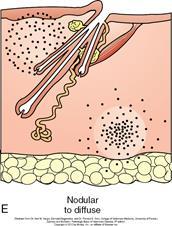
- Can have multifocal nodules – if let them go on, the will coalesce and merge.
- Depending on aetiological agent, cytokines are sent and this will lead to attraction of particular white blood cells.
- Convergence of nodules à diffuse pattern
- Cells vary:
- Neutrophils - pyogenic agents
- Histiocytes/macrophages - FBs, mycobacteria
- Neutrophils and macros – furunculosis, fungi?
- Eosinophilic – parasitic?
- Lymphocytic – insect bites, vaccine reactions
- Very common pattern in the dog.
Describe Intraepidermal vesicular/pustular dermatitis?
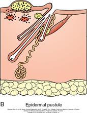
- Vesicle or pustular formation in epidermis = dermatitis(??)
- Clefting in epidermis –> vesicles or pustules
- Vesicular if just normal fluid.
- Pustular if infected, full of lots of neutrophils etc.
- Due to
- Spongiosis (intercellular oedema in epidermis)/epidermal inflammation
- parasites, infection
- Acantholysis
- infection, autoimmune disease
- Spongiosis (intercellular oedema in epidermis)/epidermal inflammation
- Intracellular oedema
- mechanical forces
The pattern of Intraepidermal vesicles/pustules is further classified by:
Position
Subcorneal (ie v superficial)
- Pemphigus foliaceus (PF), pyoderma
Suprabasilar (ie deeper)
- Pemphigus vulgaris
In follicular external root sheath
- PF
Cellular infiltrate
Neutrophils
- Bacterial pyoderma, PF
Eosinophils
- PF, parasite
What is this dog suffering from?
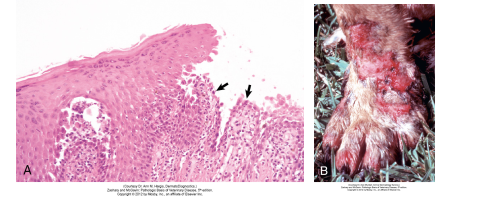
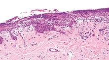
Pemphigus vulgaris, skin, dog. A, Suprabasilar clefting has left a row of basal cells (arrows) attached to the dermis. The single row of basal cells is fragile and easily damaged leading to formation of ulcers, with subsequent fluid loss and secondary bacterial infection. H&E stain.
B, Leg. Note the erythema and large confluent areas of ulceration. In contrast to pemphigus foliaceous (more commonly characterized by erosions and crusts), pemphigus vulgaris is characterized by larger more confluent ulcers because the acantholysis in pemphigus vulgaris occurs deeper in the epidermis (in the cells of the lower epidermis). Vesicles are not frequently seen as they rapidly progress to ulcers, the more common clinical lesion.
Describe Subepidermal vesicular/pustular dermatitis?
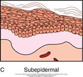
- Rare! (deeper still!)
- Entire epidermis lifts off.
- Autoimmune
- Bullous pemphigoid (see in horse on image)
- Thermal burns
- Severe dermal oedema
- Severe interface dermatitis
- but occasionally an artefact!
- Separation of epidermis from dermis
Describe Folliculitis/furunculosis/adenitis?
- Inflammation can affect various hair follicle structures, eg
- Perifollicular vascular plexus
- Perifolliculitis
- Follicular wall
- Mural folliculitis (PF, demodicosis)
- Lumen of hair follicle
- Luminal folliculitis (demodex, dermatophytes)
- Bulb
- Bulbitis (Alopecia areata)
- Sebaceous glands
- Sebaceous adenitis (auto-immune, poodle!)
- Perifollicular vascular plexus
- IF go into the wall, there is probably something in the hair follicle that attracts them and is known as luminal folliculitis.
- IF affects surrounding area and follicular wall, can cause breakdown and get release of hair into the dermis – causes massive foreign body reaction, leading to quite extensive furunculosis.
- If only cenered around bulb of hair follicle = bulbitis; specific pattern seen with alopecia areata.
- Sebaceous gland attack; quite specific attack of the gland.
- Furunculosis = perforating folliculitis with release of keratin into dermis, sets up marked inflammatory response
Describe Atrophic dermatosis?
- Not about inflammation, but instead you are losing structures of the skin! (atropy – dermis/ epidermis can get thinner, lose collagen – image showing classic case of HAC)
- Atrophy of
- Epidermis
- Hair follicles, collagen
- Sebaceous glands
- Orthokeratotic hyperkeratosis
- Follicular keratosis
- +/- calcinosis cutis if HAC
- Various endocrine causes – hormone assays to diagnose (eg HAC, hypothyroidism)
- Any chronic systemic disease or malnutrition etc.
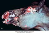
Describe panniculitis?
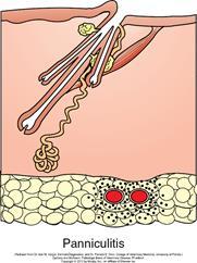
- Epidermis, follicle, dermis ok. Inflammation centred on subcutaneous fat.
- Inflammation of subcutaneous adipose tissue
- Sometimes an extension of follicular disease
- Multiple causes – histology/culture may help detect eg
- Infectious agents
- Vasculitis
- Foreign body
- Pancreatic disease
- Dog with panniculitis commonly seen in PM room with pancreatic disease. Seems to be something with the lipases.
- Often sterile idiopathic
- but must eliminate possibility of infection before treatment with corticosteroids
- Sometimes in dogs with pancreatitis or pancreatic carcinomas. Or in animals with Vitamin E deficiency or trauma
Define
Acanthocyte
Acantholysis
Acanthosis
Acanthocyte – epidermal cell free in a vesicle/pustule, caused by acantholysis
Acantholysis – loss of cohesion between cells of the living epidermis
Acanthosis – hyperplasia of stratum spinosum
Define
Apoptosis
Dyskeratosis
Epidermolysis
Apoptosis – individual cell death, requiring energy
Dyskeratosis - abnormal, premature or imperfect keratinisation of keratinocytes
Epidermolysis – degeneration of epidermal basal layer
–> separation of epidermis from dermis
Define
Exocytosis
Hydropic degeneration
Hyperkeratosis
Exocytosis – migration of inflammatory cells from dermis –> epidermis
Hydropic degeneration – vacuoles in stratum basale –> intrabasal or subepidermal clefts
Hyperkeratosis – increase in stratum corneum (orthokeratotic/parakeratotic; basket-weave/compact)
Define
Intracellular oedema
Necrolysis
Orthokeratosis
Intracellular oedema – occurs with hydropic degeneration of basal cells and ballooning degeneration (seen with herpes virus infections)
Necrolysis – epidermal necrosis with no dermal involvement and minimal inflammation
Orthokeratosis –excessive cornification – keratinocytes lose nuclei
Define
Parakeratosis
Pigment incontinence
Spongiosis
Parakeratosis - excessive cornification – keratinocytes retain nuclei
Pigment incontinence – release of melanin granules into superficial dermis
Spongiosis - intercellular oedema in the epidermis
-
CRS 1 Revision13
-
Infectious Respiratory Disease in Small Animals74
-
Bonzo has a cough..17
-
Freddie has Nasal Discharge26
-
A difficult pet show9
-
ECG revision26
-
Tracheostomy10
-
Coughing in SAs39
-
Foxy Eosinophilic bronchpneumopathy16
-
Help my dog can't breath32
-
Radiology quiz16
-
Thoracic Imaging 263
-
Thoracic imaging 338
-
Respiratory Tract Neoplasia in SA50
-
SA Airway Disease45
-
Approach to LRT Disease in SA48
-
Diseases and Conditions of the Avian Respiratory System47
-
Thoracic Radiography Quiz8
-
Lung Disease Gross Pathology25
-
SA surgery and nasal disease Wrap Up30
-
Respiratory Tract Disease in Small Animals28
-
Pleural Disease54
-
Respiratory Endoscopy35
-
Lower Airway Disease in Cats42
-
Feline Asthma (SDL)15
-
Chest Drains20
-
Sam The Dog Can't Breath6
-
Lung histopathology18
-
Small Animal respiratory parasites39
-
Blood Pressure SDL8
-
Dysrhythmias SDL41
-
SA Endocardial Disease58
-
The Ascitic Dog Cases11
-
Dysrhythmias Management25
-
Feline Cardiomyopathies52
-
Hypertension57
-
Canine Myocardial Disease53
-
Collapse, Weakness, Exercise Intolerance31
-
Cardiac Radiography50
-
Congenital Cardiac Disease48
-
Cyanosis35
-
Radiography of CRS8
-
Management of Heart Failure78
-
Heart Failure Problems10
-
Management Of Heart Failure 223
-
Echocardiograpy Images10
-
Pericardial disease in SA37
-
How to echo33
-
Pericardiocentesis5
-
Thoracic Surgery14
-
Cardiac Cases6
-
Felix11
-
AB selection for respiratory disease37
-
ECG generation14
-
ECG interpretation27
-
CPCR32
-
Triage58
-
Emergency and Critical Cases12
-
Important factors to consider in the emergency and critical care patient27
-
Traumatic Brain Injury38
-
Important factors to consider in the emergency and critical care patient COPY27
-
Nutrition for critically ill patients54
-
Anaesthesia For The Critical Patient: Is It Different?31
-
Critical patients monitoring37
-
CRS ECC Cases20
-
Formative questions46
-
Adrenal glands6
-
Pancreas4
-
Pituitary gland11
-
Thyroid and parathyroid7
-
Hyperglycaemia7
-
Canine hypoadrenocorticism30
-
Canine hyperadrenocorticism37
-
Hepatic lipidosis/ketosis/farm16
-
Feline DM34
-
Canine diabetes mellitus45
-
Diabetic Ketoacidosis27
-
Hypoglycaemia23
-
Feline hyperthyroidism37
-
Canine hypothyroidism34
-
Calcium disorders in SA25
-
Dermatology64
-
Endocrine cases29
-
PU/PD21
-
Uncommon endocrine disorders15
-
Bacterial skin disease43
-
Fungal skin disease22
-
Viral and protozoal skin disease23
-
Parasitic skin disease49
-
Autoimmune and immune mediated diseases of the skin32
-
Practical information14
-
Pattern analysis21
-
Allergic skin disease: pathophysiology and presentation18
-
Management of allergic skin disease29
-
Clinical signs of and approach to pruritus22
-
Appraoch to cutaneous masses35
-
Feline eosinophillic granuloma complexes11
-
Cytology of cutaneous masses28
-
FNA cytology practice7
-
Non-neoplastic masses21
-
Approach to alopecia27
-
Approach to pustular, papular, scaling and crusting skin disease36
-
Otitis48
-
Otitis available treatments and choice15
-
Otic surgery33
-
Dermatological therapeutic case discussion7
-
Feline specific aspects of dermatology18
-
Dermatology cases18
-
SA Anatomy Refresher17
-
Year 2 Formative (Revision)46
-
Introduction5
-
Clinical Approach To GI Disease in SA34
-
Sickly Dog35
-
Vomiting In Dogs and Cats54
-
Gastric Disease in Dogs and Cats57
-
Diagnostic Imaging of GI15
-
Practical: Dog anatomy32
-
SA GI radiography and Ultrasonography71
-
Management of SA oesophageal and gastric SA disease39
-
Pancreatitis43
-
Intro To Intestinal Disease72
-
SI Disorders in Dogs and Cats61
-
Chronic Diarrhoea in SA50
-
Management Plans for Vomiting SA16
-
Devising a Plan12
-
SA Large Intestinal Disease49
-
Why Is This Patient Vomiting Cases27
-
Approach to abdominal radiography21
-
Abdo surgery: intro and biopsy techniques38
-
Abdominal Palpation9
-
Sedation and GI plans for GI patient15
-
Abdominal radiology cases10
-
Weight loss and ascites58
-
Feline Liver Disease72
-
Liver Disease Introduction71
-
Chronic Liver disease in dogs51
-
SA Acute Liver Disease49
-
What Is Wrong With These Patients?19
-
GDV and Gastropexies54
-
Small Animal Acute Abdomen60
-
Abdominal surgery13
-
Feline infectious peritonitis40
-
Gastrotomy, Enterotomy and Enterectomy38
-
Liver, biliary tract and portosystemic shunts48
-
Surgical Diseases of The Oesophagus25
-
Anal and Rectal Conditions66
-
Hernias & Ruptures47
-
Oral tumours, oral surgery & stick injuries50
-
Rabbit Dental Disease15
-
Common Intoxications58
-
Peridontal Disease10
-
Toxiocology cases32
-
Extraction Technique28
-
Oral surgery: surgical extraction technique34
-
GI Disease in Herbivorous Rodents53
-
Formative27
-
Developmental and Genetic Dental Abnormalities9
-
Supporting the Poison Case34
-
Clinical Case10
-
Feline Oral Disease23
-
Pancreatic disease16
-
Bella and her liver16
-
Oral cavity exam33
-
Oral cavity trauma and infection32
-
Introduction COPY4
-
Erythroid110
-
Leukogram62
-
Leukaemia and Lymphadenopathy28
-
Anaemia54
-
Pathology of Lymphoid System58
-
Haemostatic Disease67
-
Diagnosis and Management of Lymphoid Diseases96
-
Immune Mediated Disease64
-
Viruses and Lymphoid System44
-
Blood Transfusions41
-
Formative assessment87
-
Revision practical16
-
Bone pathology51
-
Bone: Diagnostic Imaging70
-
SA MSK Radiography and Radiology20
-
Spine Head and Neck Imaging37
-
Autoimmunity37
-
Principles of Fracture Management 1125
-
Principles of fracture management 2100
-
Case: Bony swelling in dog16
-
NSAIDs58
-
Examination of the Lame Dog and Cat49
-
SA Joint Dx and OA51
-
Sprains and luxations46
-
Joint Treatment6
-
Decision making in Diagnostic Tests CR11
-
Antibiotics51
-
Bones and Joint Histology (and a few pics)28
-
Synoviocentesis7
-
Joint - Inflammatory Conditions60
-
SA bandaging30
-
Triage COPY19
-
Soft Tissue Trauma65
-
Radiology wrap up25
-
Orthopaedic infections46
-
CSGTR Flat Cat10
-
SA developmental disease66
-
SA Developmental Disease Part 260
-
Small Animal Distal Limb78
-
Surgical Planning2
-
Restricting Exercise8
-
SA Muscle Disease27
-
Muscle Pathology10
-
Post Op Managment60
-
SA Hindlimb51
-
SA Forelimb63
-
SA Trauma55
-
Salvage Procedures48
-
Neurodegenerative Disease33
-
Vascular Disease35
-
Neurology Infectious Diseases84
-
Ophthalmic Exam85
-
Conjunctiva and KCS60
-
Corneal Ulcers58
-
Pain Assessment17
-
Non-Notifiable Viral Disease44
-
Notifiable Neuro Viruses26
-
Neuropharmacology47
-
Eye and Disease30
-
Feline Ophthalmology32
-
Behaviour21
-
Ocular Emergencies40
-
Ocular Pharmacology81
-
Toxicities72
-
Inflammatory Disease59
-
Pain Treatment16
-
Neuropathology36
-
Neurosurgery37
-
Exotics Neurology17
-
Pharmacological Control Of Pregnancy and Parturition25
-
Formative COPY43
-
Principles of Pregnancy Failure14
-
Intro to repro pathology137
-
Principles of Cycle Manipulation32
-
Bitch and queen when to spay and why?25
-
Principles of managing dystocia45
-
Monkey (not even a monkey) at the clinic16
-
Approach to Examining the Female Tract42
-
Male Castration63
-
Principles of Contraception42
-
Principles of Neonate Care and Disorders of the Neonate37
-
Principles of therapeutics and anaesthesia in of the neonate and pregnant animal40
-
Reproductive Anaesthesia44
-
Reproductive anaesthesiaCase based seminar13
-
Approach to Examining the Male Reproductive Tract47
-
Reproductive Exam19
-
Facillitator Cases17
-
Spay practical notes28
-
The optimal mating time in the bitch22
-
Urinary Incontinence30
-
Clinical Research - Cystic Endometria Hyperplasia15
-
Diagnostic Methods in the Bitch and Queen82
-
When Will Sasha Have Her Pups?31
-
Abnormalities of pregnancy, parturition and puppies65
-
Abnormalities and Infertility in Female Small Animals45
-
Dystocia Cases31
-
Manipulation of Reproduction and Contraception45
-
Pregnancy Case studies - bitch13
-
First Opinion Reproduction Cases4
-
Unwanted Mating in the Bitch10
-
Pre-pubertal neutering14
-
Bitch Spay Complications21
-
Ovariectomy/Ovariohysterectomy34
-
Surgery of the Female Reproductive Tract34
-
Abnormalities of the Mammary Gland19
-
Peri-operative management and anaesthesia for pyometra in a dog28
-
Common Diseases and Surgery in the Male Dog and Cat37
-
Reproductive Tract Diagnostic Methods and Prostatic Disease in Males76
-
Imaging SA Repro43
-
Exotic reproduction44
-
Repro formative case 16
-
Repro formative case 24
-
Repro formative MCQ23
-
Evaluation of urinary tract disease54
-
Renal Physiology21
-
CKD & AKI26
-
Fluid Therapy9
-
Infectious Diseases7
-
Drug Choices and Therapeutics21
-
Blood Results28
-
Chronic kidney disease79
-
Acute Kidney Injury65
-
Urinary pathology98
-
Urinary Imaging112
-
Fluid therapy in urinary tract disease56
-
Diagnostic approach to polyuria and polydipsia (PUPD)48
-
Neoplasia of Urinary Tract62
-
Congenital And Neonatal Abnormalities82
-
Urinary tract infections39
-
Urinary Tract Disease in Reptiles and Birds85
-
Q and A with steve and jane3
-
Small Animal Urinary Tract Imaging Cases13
-
Interpreting Urinalysis Results48
-
Haematocrit stuff5
-
Crystal quiz9
-
Pu/Pd toolkit20
-
Lower Urinary Tract Disease60
-
Feline Lower Urinary Tract Disease, Calculi and Blocked Cats48
-
Urinary Tract Surgery11
-
Is It Incontinent?8
-
FLUTD7
-
Calculi in Dogs (and Other Pets)18
-
Hypertension/renal failure CR14
-
Practical urinary pathology14
-
FLUTD CR10
-
Contrast Radiographs16
-
Fluid Therapy In Practice36
-
Urinary Incontinence COPY32
-
Principles of urinary surgery53
-
CR Billy the dalmation18
-
Formative MCQ20
-
Formative clinical reasoning6
